Radiography techniques and latest advancements
Posted on 29/04/2024
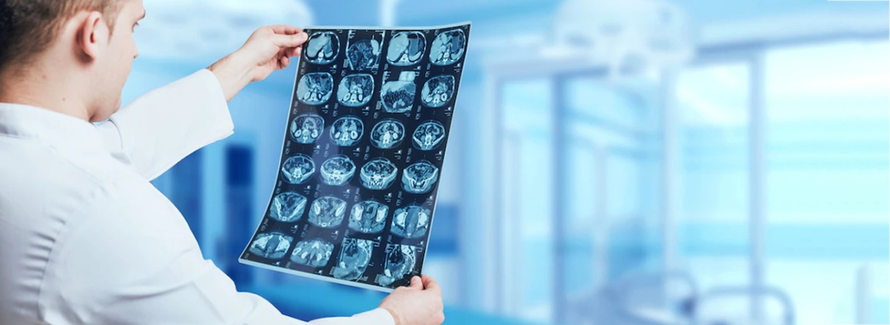
Radiography is an imaging technique using X-rays, gamma rays, or other ionizing radiation along with non-ionizing radiation to see an object's internal structure. Medical radiography ("diagnostic" and "therapeutic") and industrial radiography are two examples of radiography applications.Ionizing and non-ionizing radiation can be used to reveal the internal structure of the body on an image receptor by highlighting these differences using attenuation, or in the case of ionizing radiation, the absorption of X-ray photons by the denser substances. The body is made up of various substances with varying densities (like calcium-rich bones). Radiographic anatomy is a field of research that uses radiographic images to examine human anatomy.
There are numerous imaging modalities used in medical radiography, and each one has a unique clinical purpose.
Computed Tomography
Images produced by computed tomography, including the upper-left image in three dimensions. Ionizing radiation (x-ray radiation) and a computer are used in computed tomography, also known as a CT scan (formerly known as a CAT scan, with the "A" standing for "axial"), to produce images of both soft tissues and hard structures. Although CT utilizes more ionizing radiation than diagnostic x-rays (both of which employ X-ray radiation), levels of CT radiation exposure and scan times have decreased due to technological advancements.
X-ray absorptiometry with dual energy
Bone densitometry, often known as DEXA, is frequently used for osteoporosis examinations. As the X-rays are produced in two confined beams that scan over the patient at 90 degrees to one another, it is not projection radiography. Typically, a picture is taken of the hip , low back , or heel , and the bone density is calculated and assigned a number.
Contrast radiography
In contrast radiography, the structures of interest are visibly distinguished from their surroundings using a radiocontrast agent, a sort of contrast medium. Contrast agents are necessary for traditional angiography and may be utilized with computed tomography as well as projectional radiography (called contrast CT).
Fluoroscopy
Thomas Edison invented the term "fluoroscopy" during his early X-ray research. The method offers radiographs that move during projection. The primary purposes of fluoroscopy are to observe motion (of tissue or a contrast agent) or to direct a medical procedure like angioplasty, pacemaker implantation, or joint repair or replacement. The final procedure can frequently be completed in the operating room with the use of a C-arm, a portable fluoroscopy device. The surgical table can be moved around as it creates digital images for the surgeon. The only difference between biplanar fluoroscopy and single-plane fluoroscopy is the simultaneous display of two planes. Working in two planes is crucial for orthopedic and spinal surgery because it eliminates the need for repositioning, which can lengthen operating durations.
Recent Advancements in Radiography
Digital mobile radiography, dual-energy imaging, tomosynthesis, computer-aided diagnosis, automatic picture stitching, and AI-assisted X-ray interpretation are just a few of the recent developments in the field of digital radiography. These developments have enhanced image quality, supporting better patient outcomes and enhancing patient care.
Additionally, digital radiography has the following benefits:
- Shorter exposure times
- Improved detail detectability
- Extremely high image quality
- Faster processing and diagnosis
- Images that are easier to store and share with doctors
- Capability for remote viewing from connected digital devices
X-Ray Interpretation With AI Assistance
The field of radiology has made great progress in the analysis and interpretation of imaging data thanks to developments in computer vision, machine learning (ML), artificial intelligence (AI), and deep learning techniques. By expediting and increasing the accuracy of injury and disease diagnosis and treatment, radiology personnel can enhance the quality of patient care by using AI-assisted X-ray interpretation.
This is how AI-assisted X-ray interpretation operates:
It quickly and accurately uses algorithms to analyze data and images from both open and closed medical databases. To locate trends and abnormalities, it then compares the photos to earlier discoveries.
The following are advantages of using AI-assisted X-ray interpretation in diagnostic imaging:
- Faster tracking of crucial information for diagnosis
- The ability to prioritize critical cases better
- Reduction of errors in reading electronic health records (EHRs)
Automatic Image Stitching
Various fields use image stitching in digital radiography, which is the act of merging together smaller images to create a bigger one.A digital radiology scanning device cannot simultaneously scan the entire human body .If radiology professionals want to see a picture of a patient’s whole bone structure to look for anomalies that are indicative of potential medical issues, they can use image stitching technologies to “stitch” together images of different areas of the body.The method of combining several photographs without the assistance of a human is known as automatic image stitching.
Digital Mobile Radiography
Digital mobile radiography equipment has wheels and is designed for easy transport in healthcare settings, such as hospitals, where it can be moved to a patient’s bedside. A transportable X-ray device, for instance, can photograph patients who are immobile after surgery to let the surgeon assess the procedure's results. Digital mobile radiography equipment is portable, making disinfection easier.
Conclusion
The rapid advances in clinical radiology technology and theory have dramatically improved the diagnosis and treatment of illness and injury.The application of cutting-edge technologies in recent years has significantly helped the field of radiography as a whole. Modern digital radiography (DR) imaging equipment is quicker and easier to use, produces more accurate images, and is safer for patients, all of which contribute to better patient care.
