Significance of Ultrasound in Pregnancy
Posted on 29/04/2024
A pregnancy ultrasound is a test that images the developing baby as well as the mother's reproductive organs using high-frequency sound waves. Each pregnancy requires a different number of ultrasounds.
An ultrasound, often known as a sonogram, can aid in the monitoring of normal prenatal development and the detection of any potential issues. There are a variety of advanced ultrasounds available, including a 3-D ultrasound, a 4-D ultrasound, and a fetal echocardiogram, which is an ultrasound that examines the fetus' heart in greater detail.
When is ultrasound performed ?
Ultrasound can be performed at different stages of pregnancy, including:
First trimester ultrasounds are used to confirm that the embryo is developing inside the womb (rather than in a fallopian tube, for example), confirm the number of embryos, and calculate the gestational age and the baby's due date throughout the first three months of pregnancy.
Ultrasound is used to assess the development of fetal structures such as the spine, limbs, brain, and internal organs during the second trimester, between weeks 18 and 20. The placenta's size and placement are also examined.
After 30 weeks, a third trimester ultrasound is used to ensure that the baby is still growing at a normal rate. The placenta is examined to ensure that it is not blocking the cervix.
Different types of Ultrasound
When a more detailed image is necessary, more advanced ultrasound techniques may be used. If your doctor detects abnormalities during your standard ultrasound, these may provide the information needed to make a diagnosis
3D Ultrasound
A 3D ultrasound, unlike a standard 2D ultrasound, allows your doctor to see the fetus's width, height, and depth as well as your organs. This ultrasound is extremely useful for diagnosing any complications that may arise throughout your pregnancy. A 3D ultrasound employs a specific probe and software to create a 3D image, which is similar to a regular ultrasound. It also involves specialized technician training, thus it may not be as commonly available.
4D Ultrasound
A dynamic 3-D ultrasound is another name for a 4-D ultrasound. A 4-D ultrasound, unlike previous ultrasounds, produces a moving movie of the fetus. It gives you a clearer picture of the baby's features and movements. It also does a better job of capturing highlights and shadows. This ultrasound is carried out in the same way as regular ultrasounds, but with the addition of specialized equipment.
5D Ultrasound
HD and HD Live (also called 5D ) ultrasounds allow us to capture even clearer, sharper images. These images are more defined and have better resolution. This cutting-edge technology, often known as the "flesh tone look," enables you to see your child in a realistic perspective. This will depict the infant with a reddish or pinkish hue, as if you are seeing the child inside the womb.
Applications of ultrasound in pregnancy
Fetal echocardiography
If your doctor suspects your baby has congenital cardiac abnormalities, a fetal echocardiogram is performed. This test may be performed in the same way as a regular prenatal ultrasound, however it may take longer. It captures a detailed image of the fetus' heart, including its size, shape, and structure. This ultrasound can also show your doctor how your baby's heart is working, which can aid in the diagnosis of heart abnormalities.
Elastography
Elastography is a non-invasive medical imaging technique that helps determine the stiffness of organs and other structures in your body.
This technique can be used for detecting instances at risk for preterm delivery by measuring the cervical region's stiffness/softness.
Strain elastography and shear wave elastography are two distinct methods for cervical elastography that have been developed for quantifying the physical characteristics of the pregnant cervix.Strain elastography, determines only relative values of tissue stiffness because the applied transducer pressure is unknown, whereas shear wave elastography, is a method of ultrasound imaging based on the detection of shear wave propagation through the tissue. By using inversion algorithms, this method maps the waves into elastograms and determines stiffness of the tissue by measuring the shear modulus value.
Elastography may be able to supplement digital examination and traditional ultrasound findings on the cervical effacement process during childbirth. It might be useful for figuring out how likely it is that labor will be successfully induced.
Diagnostic uses of Ultrasound
To ensure healthy fetal and uterine/placental development, ultrasound is now used to examine the developing fetus as well as the uterus and placenta. Ultrasound imaging is used to evaluate a developing pregnancy for the following purposes at its most fundamental level:
1. Confirmation of pregnancy
2. Gestational Age - Although a term pregnancy is defined medically as lasting between 37 and 41 weeks, the general conception of a "normal" pregnancy is 40 weeks gestation. It is crucial to confirm the fetus' gestational age for a variety of reasons. One such reason is to ensure normal development, the baby's growth will be measured against recognized growth charts.Gestational age will be verified against the dates provided by the mother regarding her last menstrual period in order to confirm the due date and ensure that the baby is not delivered either too early or too late.
3. Check for Multiple Pregnancies (twins, triplets, etc.) - multiple pregnancy carries unique hazards and needs to be closely watched. In order to avoid complications quick treatment is necessary for issues such cervical incompetence and a "twin to twin transfusion".
4. Placenta issues: During pregnancy, the position of the placenta within the uterus can be crucial to the health of the developing fetus and, in some cases, the mother as well. Complications like Placenta Previa, Vasa Previa, Placenta Accreta, etc. can be identified using an ultrasound.
5. Monitor Fetal Position - Depending on the baby's position during birth (breech, transverse, cephalic, or optimum), different delivery techniques may be used.
6. Check for Congenital Anomalies - Many parents want to know if their child has any genetic or congenital issues so they may either terminate the pregnancy or get ready for the challenges that come with the specific issue.
7. Monitoring fetal growth can reveal issues with the placenta or the infant's health if the baby's growth deviates from normal expectations. In any case, the issue might need to be addressed with an early intervention.
8. Check the Level of Amniotic Fluid: The fetus produces amniotic fluid, and either be too much or too little amniotic fluid may be a sign of pregnancy-related issues that may need treatment.
Ultrasound Probe Technology
The component of the ultrasound system that contacts the patient's body is the ultrasound probe, sometimes referred to as a transducer. It houses the crystals responsible for sending and receiving acoustic pulses. A transducer is a device, like a lightbulb or a television antenna, that converts energy from one form to another. Similar to this, the probe changes sound into electrical and vice versa before sending the information to the ultrasonic device for processing and display.
Types of Ultrasound probes
Probe types are most easily determined by looking at the shape of the probe.Each sort of probe has a specific purpose, while some can be applied to a wide range of tests.
Linear Probe
Linear probes have a flat array and appearance. Piezoelectric crystals in a linear configuration are used in a linear probe to produce a straight sound wave. Linear probes have a wide range of applications, including vascular, breast, thyroid, tendon, and other examinations. These probes typically run at a high frequency and feature a rectangular beam with high near-field resolution to generate a greater image resolution.
Convex probe
Convex probes, also known as curved linear probes, contain an array that is curved, enabling a larger field of view. The convex array probe's piezoelectric crystals are made up of a curvilinear arrangement. These sophisticated acoustic probes can have more than 500 different elements in their crystal structure. These are typically excellent for in-depth analyses. These transducers are excellent for vascular, abdominal, OB/GYN, nerve, and musculoskeletal tests and have a range of uses. Convex probes are typically utilized for abdominal scans because of their design because they provide a wider and deeper view.
Endocavitary probe
Endocavitary probes have a "U" shaped lens and array in addition to a significantly longer probe handle. These probes are utilized to scan the body's inside. Endocavitary probes have a smaller depth range due to their form, but they have a wider area than even convex probes.
Phased Array / Cardiac probe
A smaller handle, square-shaped lens, and array are features of phased array or cardiac probes. They typically scan pictures of the heart. Phased array probes will be able to reach the heart and take an image because of their increased depth. This transducer's name derives from its phased array crystal configuration. Less crystals are present in phased array transducers, which causes the crystals to fire in phases to produce the image displayed on the screen.
TEE (Transesophageal) Probe
TEE probes are cardiac-type probes that require insertion into the patient's esophagus and stomach to produce an occluded image of the heart. The handle controls are used to steer these probes in one of four directions.
3D/4D Probe
With the exception of a moving array, 3D probes perform the same tasks as 2D probes. Slices of images are captured from various angles by the array inside the probe. A 3D still image or a 4D live image is created by combining all of the collected slices.
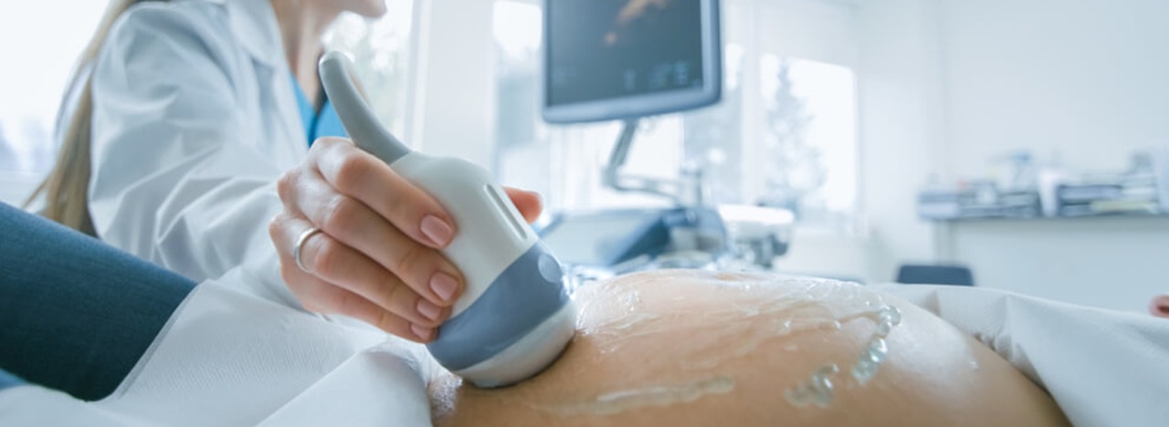
How is the procedure done?
For a trans abdominal fetal ultrasound a special gel will be applied to your abdomen by your health care practitioner or technician. This will improve sound wave transmission by eliminating air between your skin and the transducer.The transducer will be moved or scanned back and forth over your abdomen by your health care physician or technician. On a monitor, sound waves reflected off your bones and other tissues will be translated into visuals.
Your baby's anatomy will be measured by a health care provider or technician. To document essential structures, he or she might print or save select photos.
Reliability of Ultrasound
Undoubtedly, the most used diagnostic technique utilized in obstetrics is ultrasound. It is convenient, painless, produces quick, significant results, and is generally regarded as safe. Many parents view the ultrasound as a chance to glimpse their unborn child. An ultrasound can offer crucial diagnostic details about a growing child, such as verifying pregnancy and gestational age, screening for multiple pregnancies, congenital disorders, and/or placental issues, monitoring fetal position, growth, and amniotic fluid levels.
